
Pathomorphology microscopy
200 UAH
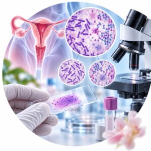
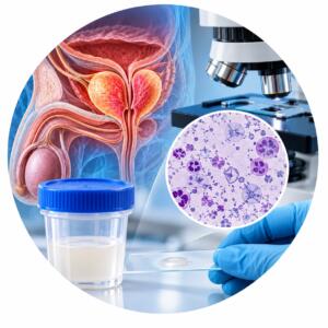
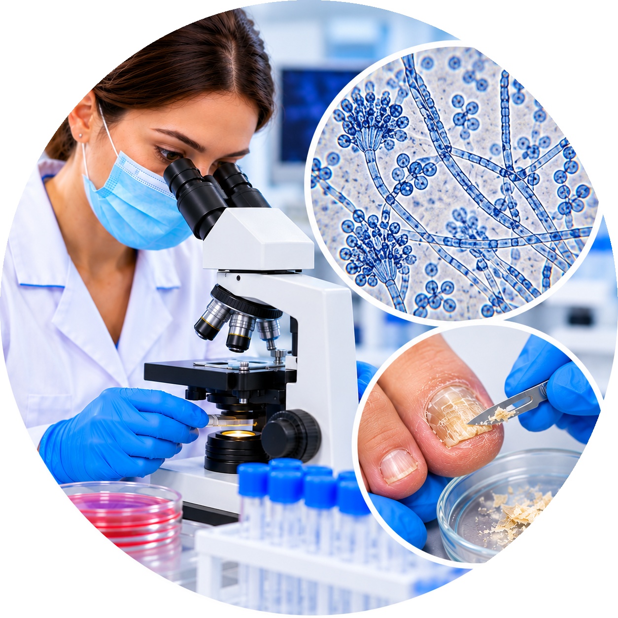
Microscopic examination of biological material to detect pathogenic fungi. The analysis helps determine the presence of fungal elements in the sample under study.
Microscopy of pathogenic fungi is an important component of the diagnosis of various diseases caused by fungi. Research using light-optical methods plays a major role in establishing the type of fungus, as well as assessing its morphological and structural features. In this article, we will consider the main aspects of microscopy of pathogenic fungi and its importance in medical practice.
Microscopy: Basics and Methods
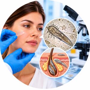
Microscopy is the science of studying objects that cannot be seen with the naked human eye, using special instruments - microscopes. Light microscopy is one of the most common methods of studying biological objects, such as cells, bacteria and, of course, fungi. The study of microorganisms using light microscopy allows us to identify their morphological characteristics and structure. For example, demodicosis mite under a microscope has a specific shape and size that helps in the diagnosis of skin diseases. It also opens up new possibilities for the study of biosystems and the interaction of living organisms.
Fungi are organisms that belong to the kingdom of fungi. They differ from other microorganisms in their cellular structure and method of nutrition. Light-optical microscopes are used to study fungi, which allow obtaining images of their structure. The study of fungi opens up new prospects in biotechnology and medicine. One of the important aspects of studying fungi is microscopy of yeast-like fungus in science, which allows for detailed analysis of their morphology and study of developmental mechanisms. This knowledge can be used to create new drugs and improve fermentation processes.
The main stages of microscopy of pathogenic fungi
- Material collection: The first step in the study of a pathogenic fungus is to collect material for analysis. Usually this is a biological sample that contains fungal structures. For example, it can be a skin swab to detect dermatophytes or a mucous membrane discharge to determine candidiasis.
- Sample preparation: The collected material undergoes processing, which may include fixation, staining, and dehydration. These procedures help preserve the structure of the fungus and improve image quality.
- Study under a microscope: After preparing the sample, it is examined under a light-optical microscope. The optical system of the microscope allows you to magnify the object and the details of the fungal structure.
- Analysis and photography: During microscopy, the structural features of the fungus are recorded and photographed for further analysis and documentation.
The value of light microscopy in the diagnosis of fungal infections
Pathogenic fungus microscopy is of great importance in medical practice. It allows:
- Identify the type of fungus: Identifying the type of fungus helps to establish a diagnosis and choose the appropriate treatment.
- Assess the extent of the damage: Microscopy allows you to determine how many fungi are in the sample, which helps to assess the extent of the lesion.
- Follow up on treatment: Repeated microscopic examinations allow to determine the effectiveness of treatment and, if necessary, to change therapy.
Conclusion
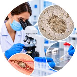
Microscopy of the pathogen using light-optical methods is an integral part of the diagnosis of fungal infections. It allows you to identify the type of fungus, determine the extent of the lesion and monitor the effectiveness of treatment. This method is an important tool for doctors and helps patients recover faster from fungal diseases. In addition, microscopy of scabies mites in medicine also plays an important role in the diagnosis of skin diseases associated with these parasites. This allows doctors to quickly identify infestations and prescribe appropriate treatment. Therefore, combining both methods increases the chances of a quick and accurate diagnosis.
| Execution time | 1 day |
|---|---|
| Type of biomaterial | Scraping the test sample |
Useful tests