
Microscopy of the scabies mite
200 UAH
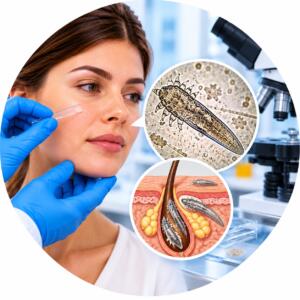
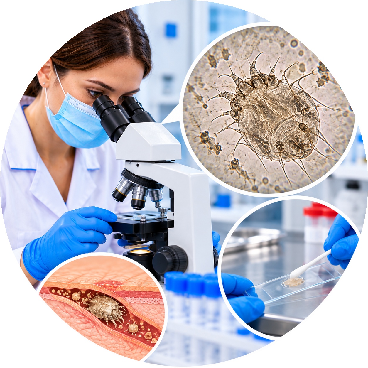
Microscopic examination of skin material to detect scabies mites Sarcoptes scabiei. The analysis helps confirm the presence of the pathogen in the test sample.
The scabies mite, or scientifically known as Trombidium, is one of many groups of arachnids that are related to the spider class. These microscopic creatures, commonly found in natural environments such as forests and gardens, play an important role in ecosystems by feeding on plant sap and other microorganisms.
To study the scabies mite and understand its morphology and life cycle, scientists often use microscopy. In this article, we will look at which light-optical methods Microscopy can be used to study scabies mites.
- Sample preparation: The first step in microscopy of scabies mites is proper specimen preparation. A fairly simple method of fixation and dehydration is usually used to preserve the mites in their natural state for further study.
- Lighting: Lighting is a key element in light microscopy. Different types of lighting are used to study scabies mites, such as LED, halogen, or gas discharge. It is important to choose the right light source to obtain clear and contrasting images.
- Objectives and lenses: Different objectives and lenses are used to maximize the magnification of the details of the scabies mite. Microscopes usually have objectives with different magnifications, allowing scientists to study different aspects of the mite's structure in detail.
- Focusing and contrast adjustment: Focusing helps to get clear images of the tick, and adjusting the contrast allows you to highlight certain details in the image. This is especially important when studying microstructures.
- Photo documentation: Cameras attached to the microscope are used to record the results of the study. This allows images of the scabies mite to be saved for later analysis and publication.
- Analysis and identification: After obtaining the images, scientists can analyze the structure of the scabies mite, identify its species, and study the features of its life cycle. This information can be useful for understanding the impact of scabies mites on ecosystems and developing methods to combat them, if necessary.
Microscopy of the scabies mite is an important tool for studying the biology and ecology of these small creatures. It allows scientists to obtain important data on the structure and function of the scabies mite, which, in turn, can contribute to the conservation and management of natural ecosystems. Studies conducted using the scabies mite microscopy can also help in studying the interactions between these parasites and their hosts. In addition, Pathophysiology of Fungus in Modern Science opens up new perspectives for studying other microscopic organisms and their role in nature. This allows scientists to gain a deeper understanding of the complex ecological relationships and mechanisms of adaptation of organisms.
| Execution time | 1 day |
|---|---|
| Type of biomaterial | Scraping the test sample |
Useful tests