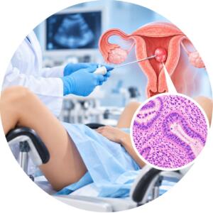


Examination of the uterus with appendages (fibroids)
800 UAH
Pathohistological examination of the uterus with appendages (for myoma) — a comprehensive morphological study of the removed surgical material, which includes the body of the uterus with myomatous nodes, the cervix, as well as the ovaries and fallopian tubes.
Why is it prescribed: to assess the nature of myomatous nodes (histological type, growth activity, presence of dystrophic changes or necrosis), detailed study of the condition of the endometrium and cervix, as well as exclusion of malignant processes (leiomyosarcoma, endometrial cancer) and pathologies in the uterine appendages (cysts, salpingitis, ovarian tumors).
Material for research: surgical material (removed uterus along with cervix, fallopian tubes and ovaries).
Method: pathomorphological (histological) examination with histological examination and microscopy of sections from various parts of the organ and appendages.
Histological material is taken only in medical institutions.
Histological examination of the uterus with appendages — is a comprehensive pathomorphological analysis of surgical material (uterus, fallopian tubes and ovaries) removed during hysterectomy for fibroids (leiomyomas). The study is a mandatory stage after surgery to confirm the benignity of the nodes and assess the condition of all removed organs.
Research objective: The main task is to verify the diagnosis of myoma and exclude leiomyosarcoma (a malignant tumor of the muscle layer). Simultaneous examination of the appendages (tubes and ovaries) allows to detect concomitant pathologies, such as cysts, chronic inflammations or hidden neoplastic processes.
What does the pathologist evaluate:
The structure of myomatous nodes: determining the type of myoma (cellular, symplastic, with degenerative changes, etc.).
Endometrium: the condition of the uterine mucosa for the presence of hyperplasia or polyps.
Myometrium: assessment of the muscle layer for signs of adenomyosis (uterine endometriosis).
Condition of the ovaries and tubes: morphological analysis of ovarian tissues (presence of follicles, cysts) and patency/changes in the fallopian tubes. Assessment of the condition of the ovaries and fallopian tubes is an important stage in reproductive diagnostics. For a more detailed study of possible pathologies, such as endometriosis or other diseases, a cervical biopsy for diagnosis. This will help establish the correct diagnosis and determine the next steps in treatment.
Cervix: exclusion of dysplasia or other pathological changes in the cervical canal.
Clinical significance: The final histological conclusion allows the doctor to confirm the radicality of the intervention and determine the tactics of further observation of the patient. This is important for understanding the general hormonal background of the body and predicting the health of the woman in the postoperative period. It is also important to consider that the results of the histological examination may affect the choice of treatment methods, such as excision and conization of the cervix. These procedures may be necessary to further eliminate any potential risks of developing cancer. Thus, timely diagnosis and adequate intervention can significantly improve the patient's health prognosis.
When is it prescribed:
after surgical treatment of large uterine fibroids;
with multiple nodes with suspected rapid growth;
in cases of combination of fibroids with ovarian or endometrial pathology;
with pain syndrome and abnormal bleeding, which led to the removal of the organ with appendages.
| Execution time | 2 days |
|---|---|
| Type of biomaterial | Tissues and organs |
Useful tests