

Examination of the ovaries and fallopian tubes
700 UAH
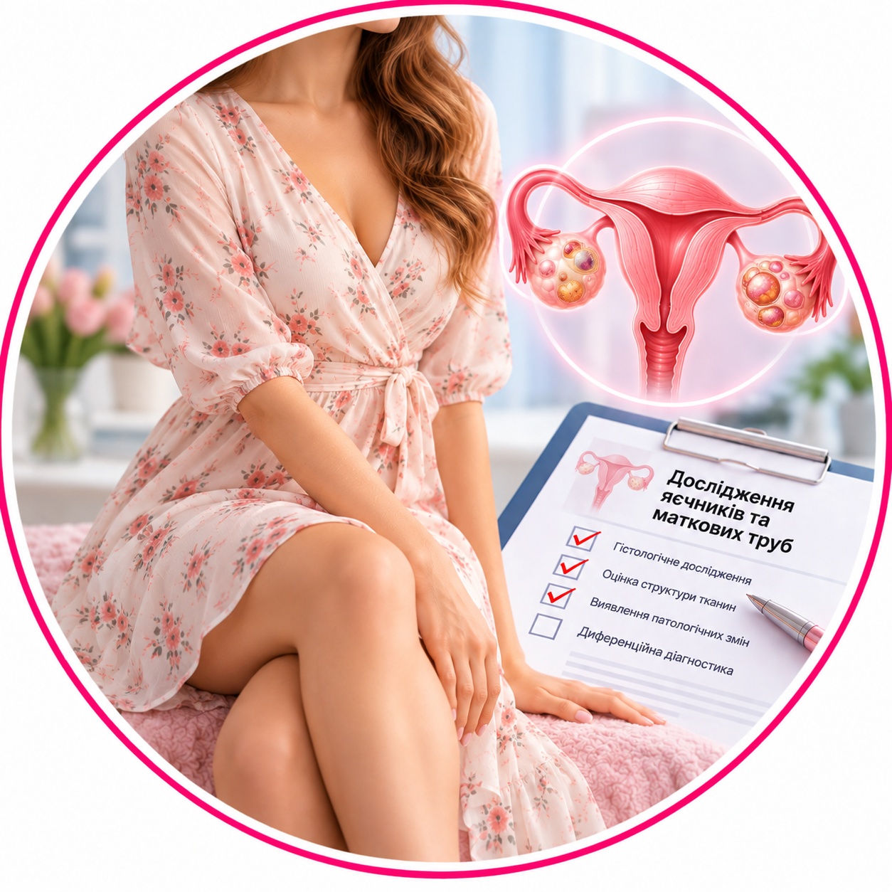
Pathohistological examination of the ovaries and uterine pipes — comprehensive morphological study of removed tissues of the female reproductive system (ovaries and fallopian tubes) to establish an accurate diagnosis and assess the nature of the detected changes.
Why is it prescribed: for histological confirmation of benign or malignant tumors (cysts, cancer), detection of inflammatory processes (oophoritis, salpingitis, adnexitis), causes of infertility, assessment of tissue condition in endometriosis or after planned removal (for example, during sterilization or as a stage of more complex operations).
Material for research: surgical material (one or both ovaries and fallopian tubes, or their fragments).
Method: pathomorphological (histological) study with the preparation and analysis of serial micropreparations.
Histological material is taken only in medical institutions.
Histological examination of the ovaries and fallopian tubes — is a comprehensive analysis of the uterine appendages removed during surgery (adnexectomy). The study allows for a complete morphological assessment of the organs, to establish the nature of pathological changes, and to rule out the presence of oncological processes.
Research objective: The analysis is aimed at a detailed study of the structure of tissues to verify benign formations (cysts, fibroids), inflammatory conditions, or detect malignant tumors. Because the ovaries and tubes are closely are anatomically related, their joint study gives a holistic picture of the spread of pathology in the pelvis.
What does the pathologist evaluate:
Ovarian tissue: type of cysts (endometrioid, serous, teratomas), presence of borderline or malignant tumors, condition of the stroma.
Fallopian tubes: patency of the lumen, presence of signs of chronic inflammation (salpingitis), signs of tubal pregnancy or hydrosalpinx.
Epithelial layer: search for atypical cells, especially in the fimbrial part of the tube, where oncological changes often begin.
Vascular network: assessment of blood circulation and the presence of possible vascular neoplasms.
Clinical significance: The histological conclusion is key to determining further tactics: the need for hormonal therapy, the amount of additional treatment by an oncologist, or simply dynamic observation. This study confirms the radicality of the operation and helps predict the general health of the woman.
When is it prescribed:
removal of the appendages in case of tumors or large ovarian cysts;
chronic inflammatory diseases of the appendages with the formation of tumor-like masses;
suspected ectopic pregnancy;
performing surgery on menopausal patients to prevent cancer risks.
| Execution time | 2 days |
|---|---|
| Type of biomaterial | Tissues and organs |
Useful tests