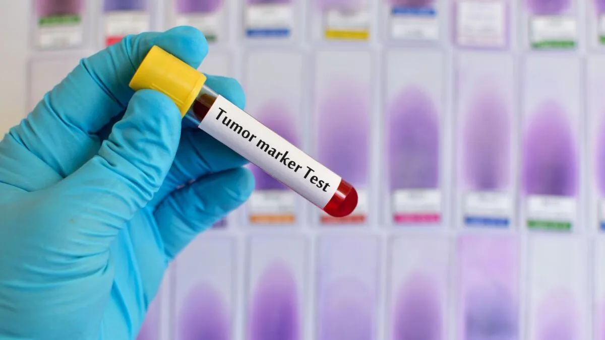
Tumor markers in liver cancer: diagnosis, significance and limits of effectiveness
The role of tumor markers in the detection of hepatocellular carcinoma
Tumor markers are biological molecules produced by tumor cells or the body in response to tumor growth. In the case of liver cancer, particularly hepatocellular carcinoma (HCC), tumor markers allow for early detection of the disease, assessment of the risk of metastasis, and monitoring of treatment effectiveness or possible recurrence. Despite their limitations in accuracy, they remain an important component of laboratory diagnostics.
The most common tumor markers in liver cancer
Alpha-fetoprotein (AFP)
This is the main and most frequently used tumor marker for HCC. AFP is produced in the fetal liver, but its level decreases after birth. In adults, elevated AFP levels indicate malignant processes, especially at levels above 400 ng/ml. At the same time, its specificity is limited: not all forms of liver cancer are accompanied by high AFP levels, and false-positive results are possible in chronic hepatitis or cirrhosis.
AFP-L3 (alpha-fetoprotein fraction)
This glycoprotein fraction is more specific for hepatocellular carcinoma, as it is predominantly synthesized by tumor cells. Elevated AFP-L3 levels even with normal total AFP may signal early tumor development.
Des-gamma-carboxy prothrombin (DCP, or PIVKA-II)
This marker is a modified form of prothrombin. Its concentration is increased in patients with HCC and it is less sensitive to cirrhosis or hepatitis compared to AFP. DCP is often used in Japan as a key tumor marker for monitoring tumor progression.
Glypican-3 (GPC3)
A protein expressed by hepatocellular carcinoma cells. It is not found in normal adult liver but is frequently detected in HCC tissues, especially in cases with normal AFP levels. GPC3 is promising as an immunohistochemical marker and potential target for therapy.

Evaluating the effectiveness of markers
In clinical practice, a panel of tumor markers is often used to improve diagnostic accuracy. For example, simultaneous assessment of AFP, AFP-L3, and DCP levels provides a more sensitive result than using a single marker. This is especially important for patients with chronic liver disease, where the risk of tissue transformation into malignancy is higher.
Table: Main tumor markers in liver cancer
| Tumor marker | Norm | The increase is characteristic of | Limitation |
|---|---|---|---|
| AFP | <10 ng/ml | Liver cancer, teratomas, cirrhosis | Possible false positives for hepatitis |
| AFP-L3 | <10% | Early stage HCC | Not found in all types of tumors |
| DCP (PIVKA-II) | <40 mAU/mL | FCC | Not suitable for patients with coagulopathies |
| GPC3 | – | Immunohistochemical confirmation of HCC | Not detected in blood serum |
Use of tumor markers in the clinic
Tumor markers in cancer liver is an auxiliary tool. None of them can definitively establish the diagnosis without the results of imaging methods (ultrasound, CT, MRI) or histology. However, in cases of suspected HCC in patients with cirrhosis, regular screening is AFP and DCP allows for the detection of tumors at a preclinical stage, when radical treatment is still possible.
Highly qualified cytomorphologist with over 45 years of experience. For 33 years, she headed the cytology laboratory of the regional oncology clinic. Expert in early diagnosis of malignant neoplasms at Medi Lab Plus LLC. More details…