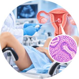


Examination of the vulva during vulvectomy
700 UAH
Pathohistological examination of vulva tissues during vulvectomy — comprehensive morphological study of the surgical material (removed vulva tissues) to determine the exact diagnosis, histological type of the tumor, the degree of its malignancy, and assess the radicality of the surgical treatment performed.
Why is it prescribed: in vulvar cancer, severe degrees of dysplasia (VIN II–III), severe forms of kraurosis/lichen sclerosus, as well as after partial or total vulvectomy to assess the resection margins (cleanliness of surgical margins).
Material for research: surgical material (fragment or completely removed vulva together with subcutaneous fat and, if necessary, lymph nodes).
Method: pathomorphological (histological) study with serial tissue sections.
Histological material is taken only in medical institutions.
Histological examination in vulvectomy — is an extended pathomorphological analysis of the entire volume of tissues of the external genital organs removed during surgery. This study is a critically important stage in the treatment of oncological or precancerous diseases, as it provides definitive information about the nature of the tumor and the radicality of the surgery performed.
Research objective: The main task is a complete morphological verification of the pathological process. The pathologist not only confirms the diagnosis, but also describes in detail the extent of the disease, which is necessary to determine further treatment tactics (radiotherapy, chemotherapy or observation).
What does the pathologist evaluate:
Type and tumor differentiation: definition the histological type of cancer (most often squamous cell) and the degree of its aggressiveness.
Depth of invasion: measuring how deeply the tumor has grown into the underlying tissues.
Resection margins: checking whether tumor cells remain at the edge of the incision (whether the operation is radical - "clean margin").
Presence of accompanying changes: diagnosis of underlying processes such as lichen sclerosus or vulvar dysplasia (VIN).
Lymphovascular invasion: checking for the presence of cancer cells in the vessels, which indicates the risk of metastasis.
Clinical significance: The conclusion after vulvectomy is a crucial document for the oncologist. Accurate data on the size of the tumor and the condition of the resection margins allow minimizing the risk of recurrence and developing an individual rehabilitation plan for the patient.
When is it prescribed:
after radical or partial vulvectomy for confirmed vulvar cancer;
treatment of severe degrees of dysplasia (VIN III) occupying a large area;
recurrent dystrophic diseases with suspected malignancy;
the presence of large neoplasms that require complete removal of vulva tissues.
| Execution time | 2 days |
|---|---|
| Type of biomaterial | Tissues and organs |
Useful tests