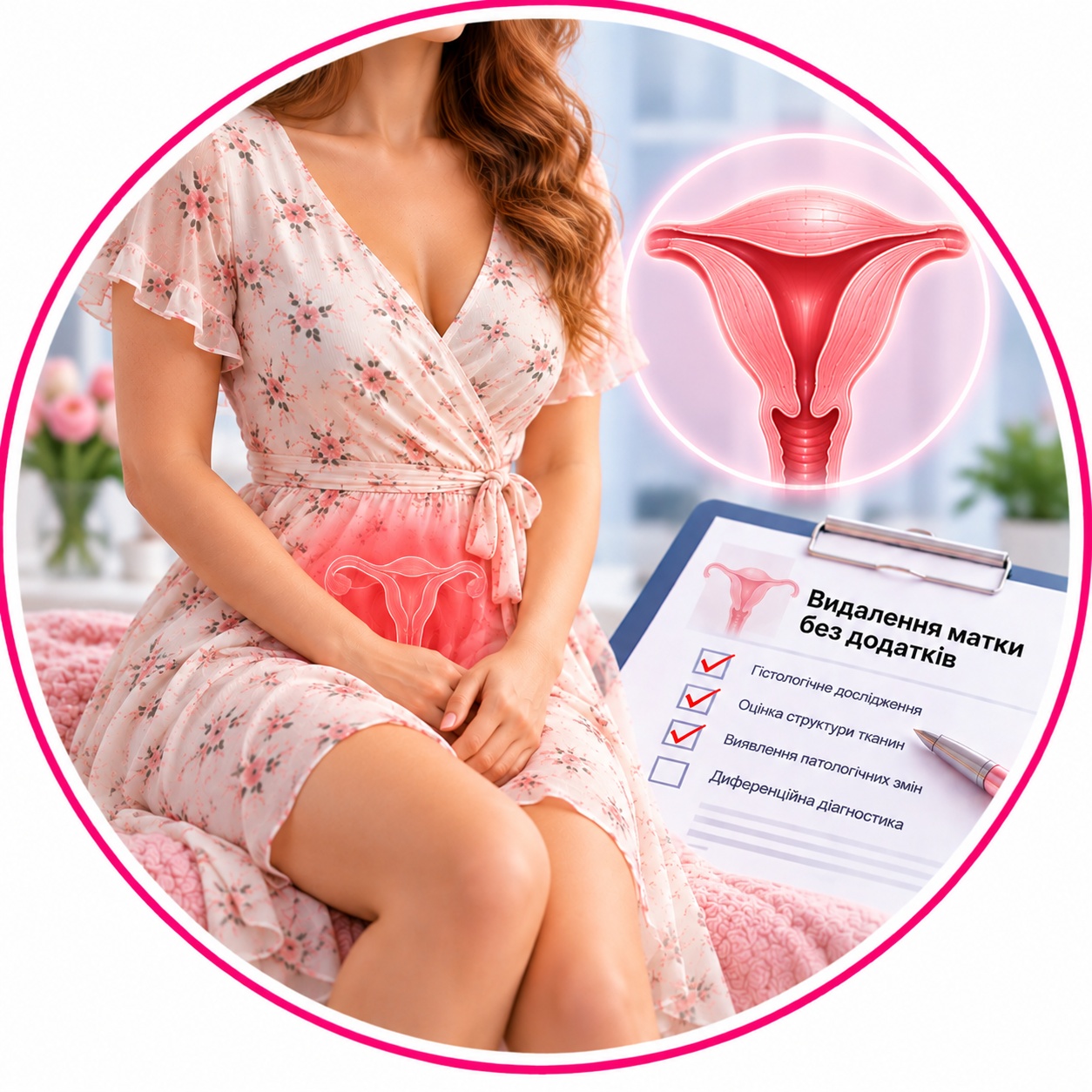

Removal of the uterus without appendages
800 UAH
Pathohistological examination of the uterus without appendages (after hysterectomy) — comprehensive morphological study of the removed body and cervix (or only the body of the uterus in case of subtotal hysterectomy) without ovaries and fallopian tubes to establish a final diagnosis.
Why is it prescribed: for multiple or large uterine fibroids, severe adenomyosis, abnormal uterine bleeding, recurrent endometrial hyperplasia, as well as to exclude or confirm malignant neoplasms (cancer of the body or cervix).
Material for research: surgical material (removed uterus without ovaries and fallopian tubes).
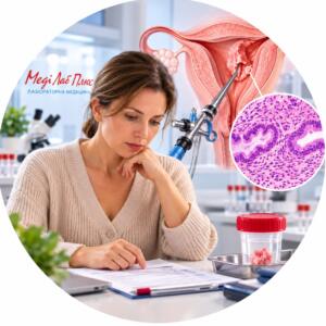
Method: pathomorphological (histological) examination with dissection of the organ and preparation of serial sections from different areas (endometrium, myometrium, cervix).
Histological material is taken only in medical institutions.
Histological examination of the uterus without appendages — is a comprehensive analysis of the surgical material (corpus and cervix) removed during subtotal or total hysterectomy. This is a mandatory stage after surgery, which allows you to obtain a complete morphological picture of the disease and confirm the benignity of the removed tissues.
Research objective: The main task of the analysis is the final verification of the clinical diagnosis. The study allows to study in detail the structure of myomatous nodes (if they were the reason for the operation), to assess the condition of the endometrium and to exclude hidden oncological processes that could not be diagnosed at the preoperative stage.
What does the pathologist evaluate:
Myometrium (muscle layer): presence and type of leiomyoma nodes, exclusion of signs of sarcoma, diagnosis of adenomyosis (internal endometriosis).
Endometrium (mucous membrane): detection of hyperplastic processes, polyps or atypical changes.
Cervix (if removed): assessment of the condition of the epithelium of the cervical canal and vaginal part for the presence of dysplasia (CIN) or cancer. The assessment also includes uterine fibroid research, if present, as fibroids can affect the overall health of the reproductive system. It is important to identify possible abnormalities in order to prescribe appropriate treatment in a timely manner. In addition, regular examinations help monitor changes in the epithelium and ensure early detection of potential pathologies.
Vascular component: assessment of the condition of the uterine vessels (presence of sclerotic changes or thrombosis).
Clinical significance: The result of the study answers the main question: was the pathology benign? This determines the further tactics of patient management - from the usual annual follow-up to the need for specific therapy from an oncologist or endocrinologist.
When is it prescribed:
after surgical treatment of uterine fibroids;
in severe forms of adenomyosis (uterine endometriosis);
with recurrent endometrial hyperplasia;
in cases of uterine prolapse, which required its removal.
| Execution time | 2 days |
|---|---|
| Type of biomaterial | Tissues and organs |
Useful tests